Dipole
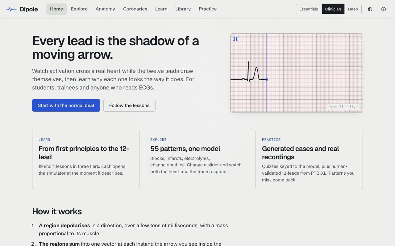
ECG atlas where a 3D dipole model draws all twelve leads.
by Talal WarsiAdded to the directory Nov 3, 2026For education, not clinical use
About Dipole
Regions of the heart depolarise, sum into a heart vector, and each lead draws its projection. There are 19 lessons, 55 adjustable patterns covering blocks, infarcts, electrolytes and channelopathies, and quizzes that mix generated cases with PTB-XL recordings.
What it does
- 19 lessons in three tiers
- 55 adjustable patterns
- Anatomy and coronary views
- Quizzes with generated cases and PTB-XL recordings
- Progress saved in your browser
Related tools
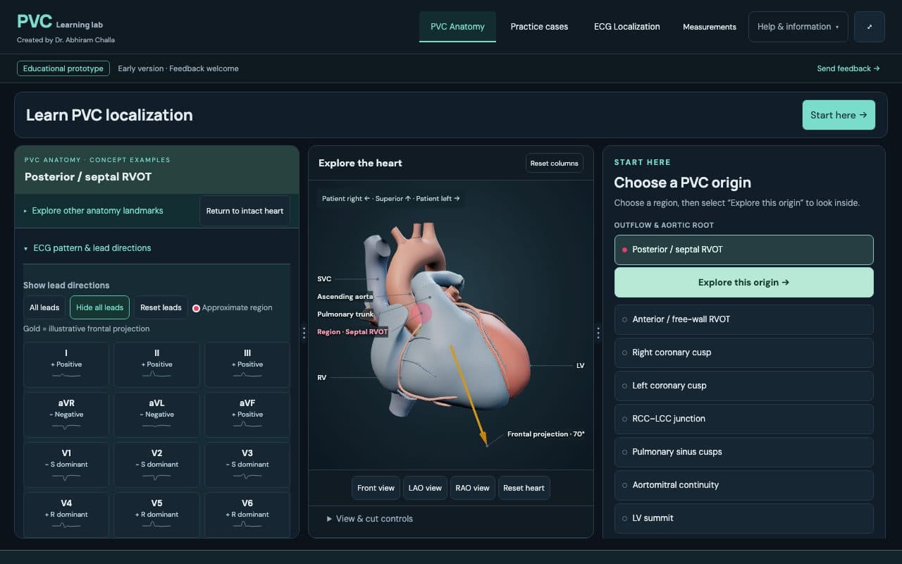
Learn PVCs
by Abhiram Challa
Learn to localize PVCs from the 12-lead using 3D cardiac anatomy.
Electrophysiology+1Free, no sign-up
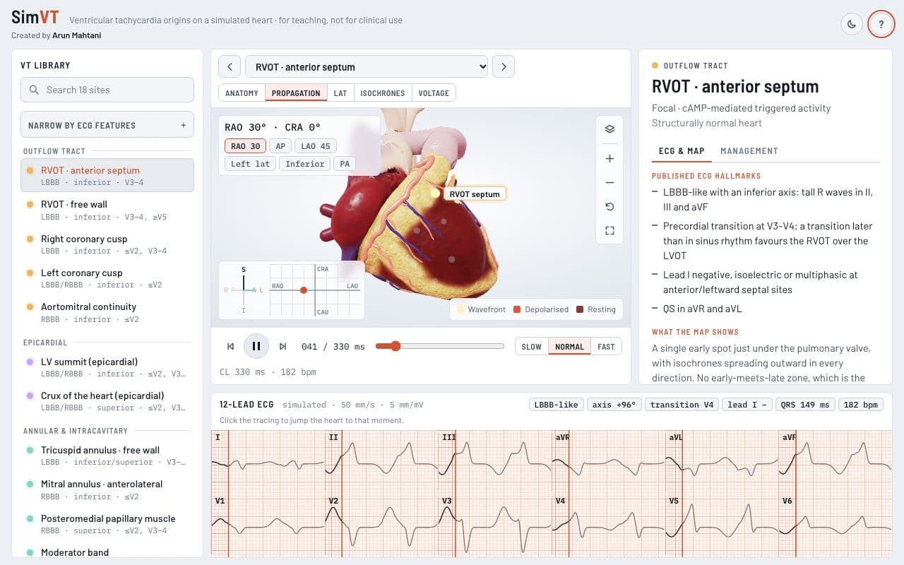
SimVT
by Arun Umesh Mahtani
VT localization on a 3D heart with a synced 12-lead ECG.
Electrophysiology+1Free, no sign-up

CardioRender
by Dinesh Raja Palanisamy
Browser 3D simulators for coronary anatomy, conduction, echo and EP.
Cardiology+2Free, no sign-up